Fixation stability
The Fixation Stability module uses the Tobii eye tracking device to measure fixation "disparity", phoria and fixation stability.
Running the Fixation Stability module
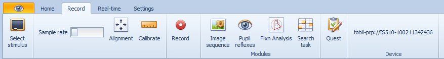
To run the Fixation Analysis module, select the Record tab at the top of the control screen and select Fixn Analysis.
This will display the Fixation Analysis Module dialog to be displayed as shown below:
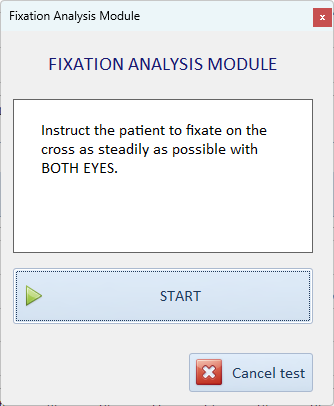
To start the test select START.
A cross will be displayed in the centre of the patient's screen.
Follow the Instructions on the screen and instruct the patient to fixate the cross as steadily as possible with BOTH EYES.
A tone will indicate the beginning and the end of the recording.
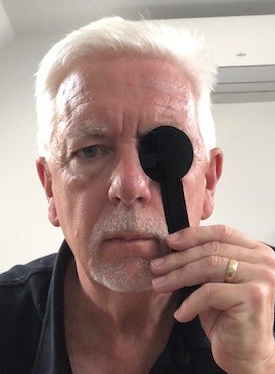
Follow the instructions on the screen and cover the patient's LEFT eye using the special IR transparent occluder included with the Clinical Eye Tracker 5L.
Select CONTINUE to start the next phase of the recording and instruct the patient to fixate on the cross as steadily as possible throughout the recording.
A tone will indicate the beginning and the end of the recording.
Follow the instructions on the screen and cover the patient's RIGHT eye using the IR transparent occluder.

Select CONTINUE to start the next phase of the recording and instruct the patient to fixate on the cross as steadily as possible throughout the recording.
At the end of the recording, select Save to add the results to the database.
To display the Fixation Analysis report, select Modules ... Fixation stability and select the result from the list.
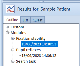
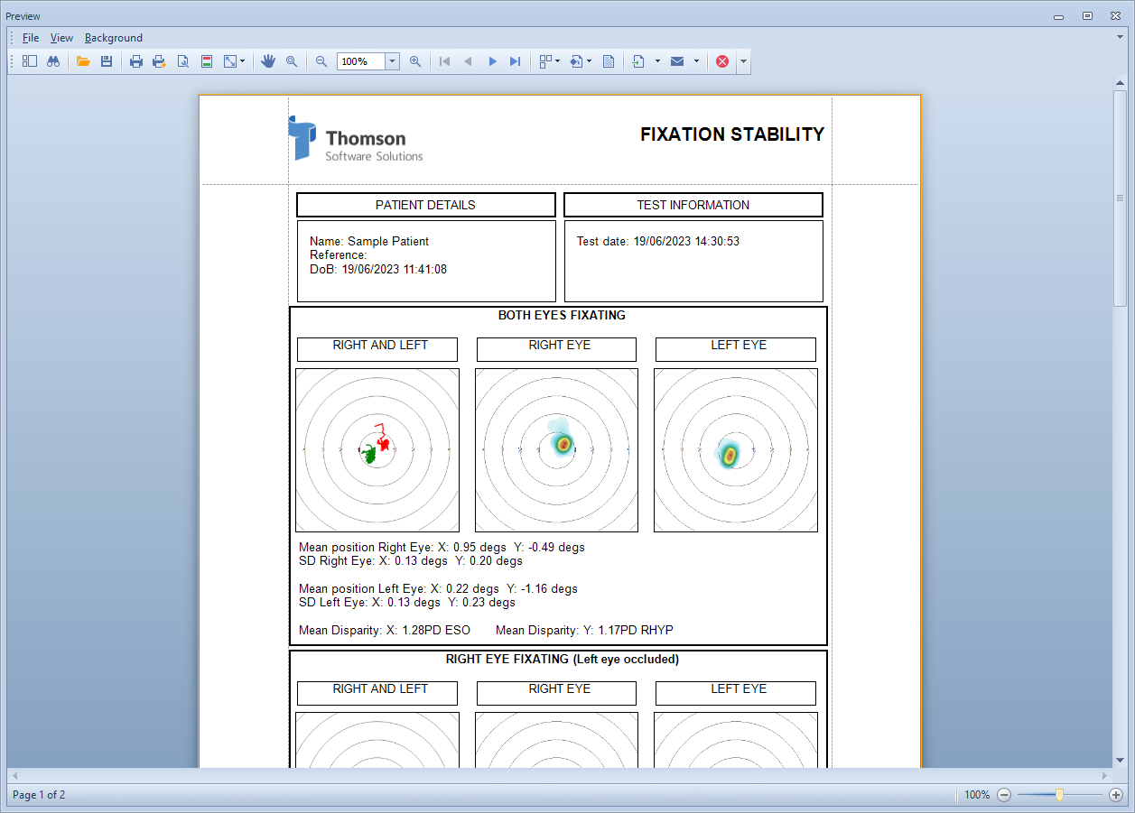
Selecting the Fixation button on the main toolbar will show the Fixation Analysis report.
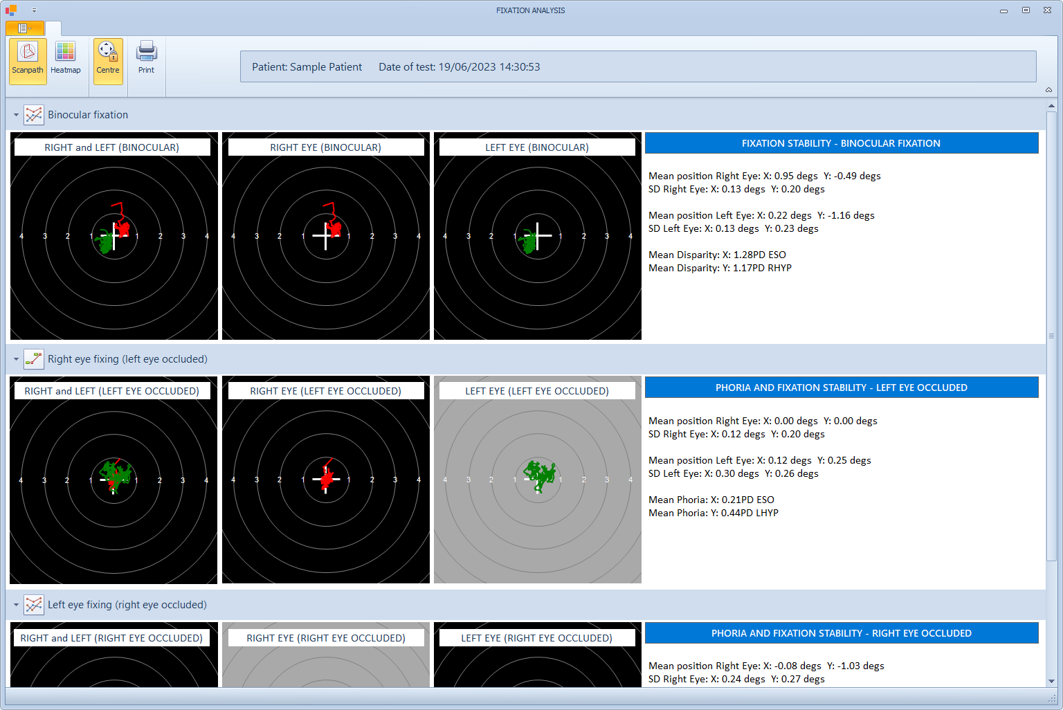
The first panel show the results with Binocular fixation. The second and third panels show the results with the Right eye fixing (left eye occluded) and the Left eye fixing (right eye occluded) respectively.
Each panel shows three XY displays showing a) the traces for each eye (RIGHT - red, LEFT - green), b) the trace for the RIGHT eye only and c) the trace for the LEFT eye only.
The concentric circles represent 1 degree intervals from the target location.
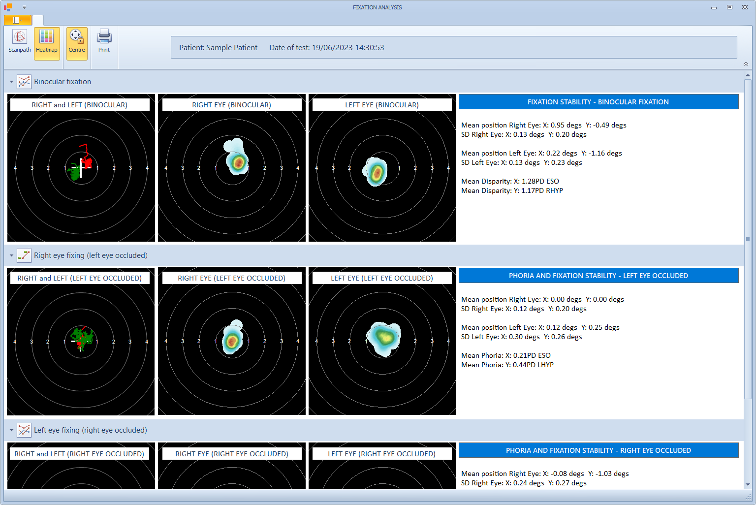
The eye position can be represented as a scan-path (as shown above) or a heat-map (as shown below), by selecting the corresponding buttons on the toolbar.
Centre traces.
The accuracy of the gaze location recorded by the Tobii Eye Tracking device is dependent on the calibration process. If the calibration data is inaccurate, the gaze location may be offset from the actual target location.
To circumvent this issue, the average gaze location is calculated for each eye using the recordings obtained when that eye was fixating (an the other eye was occluded).
If the Centre is turned on (button in toolbar), this average gaze location is taken as the "zero" position for each eye and all other results are offset by this amount. This will mean that when centering is turned on, the Mean position Right Eye and the Mean position left eye will always be zero for the Right eye fixing and the Left eye fixing panels.
Centre is turned on by default.
Binocular fixation
As the patient was binocular for this condition, any deviation between the fixation locations of the two eye must reflect a fixation disparity (or calibration error).
The information panel gives the following information:
Mean position Right Eye: The mean X and Y fixation position of the RIGHT EYE relative to the target position
SD Right Eye: The Standard Deviation (X and Y) of the fixation position of the RIGHT EYE relative to the target position. This provides a good measure of the stability of fixation of the RIGHT EYE.
Mean position Left Eye: The mean X and Y fixation position of the LEFTT EYE relative to the target position
SD Left Eye: The Standard Deviation (X and Y) of the fixation position of the LEFTT EYE relative to the target position. This provides a good measure of the stability of fixation of the LEFT EYE.
Mean Disparity X: The mean disparity in horizontal fixation position (expressed in Prism Dioptres)
Mean Disparity Y: The mean disparity in vertical fixation position (expressed in Prism Dioptres)
NOTE - While this disparity reflects a difference in fixation location while the patient is binocular, it is not equivalent to a conventional clinical measurement of fixation disparity (where the amplitude of the prism required to align nonius lines is used as the measure of fixation disparity).
Right eye fixing (left eye occluded) and Left eye fixing (right eye occluded)
As the patient had one eye occluded for these conditions, any deviation between the fixation locations of the two eye must reflect a phoria (or calibration error).
The information panel gives the following information:
Mean position Right Eye: The mean X and Y fixation position of the RIGHT EYE relative to the target position
SD Right Eye: The Standard Deviation (X and Y) of the fixation position of the RIGHT EYE relative to the target position. This provides a good measure of the stability of fixation of the RIGHT EYE.
Mean position Left Eye: The mean X and Y fixation position of the LEFTT EYE relative to the target position
SD Left Eye: The Standard Deviation (X and Y) of the fixation position of the LEFTT EYE relative to the target position. This provides a good measure of the stability of fixation of the LEFT EYE.
Mean Phoria X: The mean difference in horizontal fixation position (expressed in Prism Dioptres)
Mean Phoria Y: The mean difference in vertical fixation position (expressed in Prism Dioptres)
Printing a report
A bespoke report show the Fixation stability results can be created by selecting Print from the toolbar.